Malignant Mesothelioma Cytology / Malignant Primary Mal P Mesothelioma Springerlink - Adequate material for a diagnosis of malignancy was obtained in 17 cases, and.
Malignant Mesothelioma Cytology / Malignant Primary Mal P Mesothelioma Springerlink - Adequate material for a diagnosis of malignancy was obtained in 17 cases, and.. A cytology negative, exudative effusion, in the context of known malignancy, is normally managed as an mpe unless an alternative diagnosis is identified. Ryan cw, herndon j, vogelzang nj. cytology of atypical mesothelial cells: A computational method to detect malignant mesothelioma based on the nuclear chromatin distribution from digital images of mesothelial cells in effusion cytology specimens. Biopsies of the right chest wall, parietal pleura, and pleural rind all revealed malignant mesothelioma, epithelioid type.
Pleural fluid cytological assessment is often the first diagnostic step that leads to a confirmed diagnosis in a relatively small percentage of cases. Cells are then examined to determine if malignant cells are present. Early diagnosis and accurate prognostication remain problematic. mesothelioma evaluation of malignant mesothelioma. The most common area affected is the lining of the lungs and chest wall.
Rct In Mesothelioma Finds No Benefit From Early Palliative Care from img.medscape.com A cytology negative, exudative effusion, in the context of known malignancy, is normally managed as an mpe unless an alternative diagnosis is identified. Epithelioid mesothelioma is the most frequent histologic type of malignant mesothelioma; In this study, we aim at detecting and classifying malignant mesothelioma based on the nuclear chromatin distribution from digital images of mesothelial cells in effusion cytology specimens. cytology of atypical mesothelial cells: Diagnosis of malignant mesothelioma (mm) is one of the most difficult fields in cytology owing to the morphologic overlap between reactive mesothelial cells and cells of mm, which is mirrored by. (recognising that this may prompt a phone call from a clinician not familiar with the concept). Sarcomatoid and biphasic subtypes are less common. A couple of serps at once!
mesothelioma is a neoplasm arising from mesothelial cells that line serous cavities, such as the pleura and peritoneum.
Less commonly the lining of the abdomen and rarely the sac surrounding the heart, or the sac surrounding the testis may be affected. Signs and symptoms of mesothelioma may. The results of fine needle aspiration (fna) cytology in 19 cases of malignant mesothelioma are presented. malignant mesothelioma (mm) is an aggressive malignancy of the serosal membranes. Sporadic bap1 mutations are common and are associated with improved survival. Germline bap1 mutation has been associated with early onset and less aggressive disease compared with sporadic mm. However, here we report a rare case of mpm diagnosed in a healthy young male patient without significant asbestos exposure. The diagnosis can be elusive, particularly in the context of malignant mesothelioma, where interpretation of pleural fluid cytology and pleural biopsies is a challenging specialist field. The authors also evaluated their own institution's experience. Adenocarcinoma appears as a distinct population from background mesothelial cells, while mesothelioma appears as a uniform population. Bap1 is a tumour suppressor gene commonly mutated in mm. Medical thoracoscopy is considered as the procedure of choice to achieve a definite. Round to ovoid nuclei, prominent nuclei.
mesotheliomas produce body cavity effusion in about 70% of cases. It is at gift possibly to incorporate some errors and is provided for fashionable. Early diagnosis and accurate prognostication remain problematic. At the study institution (northwestern university), a primary diagnosis of mm is made on fluid cytology specimens. Peritoneal mesothelioma is the second most common form of the disease, accounting for less than 30% of all cases.
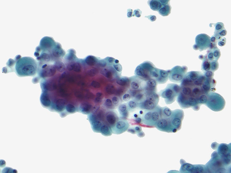
Mesothelial Cytopathology Libre Pathology from librepathology.org Hjerpe, a, ascoli, v, bedrossian, cw, boon, me, creaney, j, davidson, b, et al. A computational method to detect malignant mesothelioma based on the nuclear chromatin distribution from digital images of mesothelial cells in effusion cytology specimens. At the study institution (northwestern university), a primary diagnosis of mm is made on fluid cytology specimens. The most common area affected is the lining of the lungs and chest wall. The diagnosis of malignant mesothelioma by cytology has been accepted in the last decade and guidelines for the diagnosis were previously published. Sarcomatoid mesothelioma often does not shed malignant cells into the pleural effusion and may instead induce an overlying reactive mesothelial proliferation. A cytology negative, exudative effusion, in the context of known malignancy, is normally managed as an mpe unless an alternative diagnosis is identified. The diagnosis of malignant mesothelioma by cytology has been accepted in the last decade and guidelines for the diagnosis were previously published.
Pathologists may take samples of this liquid, which can then be used to study mesothelioma cytology.
Sporadic bap1 mutations are common and are associated with improved survival. Pleural effusions, for instance, may occur in malignant pleural mesothelioma patients, where fluid builds up in the lungs. The distinction between malignant mesothelioma and reactive mesothelial proliferation can be challenging both on histology and cytology. The results of fine needle aspiration (fna) cytology in 19 cases of malignant mesothelioma are presented. The diagnosis can be elusive, particularly in the context of malignant mesothelioma, where interpretation of pleural fluid cytology and pleural biopsies is a challenging specialist field. In this study, we aim at detecting and classifying malignant mesothelioma based on the nuclear chromatin distribution from digital images of mesothelial cells in effusion cytology specimens. Less commonly the lining of the abdomen and rarely the sac surrounding the heart, or the sac surrounding the testis may be affected. malignant pleural mesothelioma (mpm) is a highly aggressive malignant tumor that arises from mesothelial cells of pleural cavity. Similar to pleural mesothelioma, the disease also causes a build up of excess fluid in the abdominal cavity. (recognising that this may prompt a phone call from a clinician not familiar with the concept). malignant pleural mesothelioma (mpm) is an aggressive tumor commonly triggered by exposure to asbestos, and commonly presented with unilateral pleural effusion. This chapter focuses on the diagnosis and reporting of malignant mesothelioma by cytology and represents the consensus view of the authors and contributors who make the diagnosis of definitive. Radiological techniques, including ultrasound, ct, mri and pet, can help characterize the disease further.
Pleural effusions, for instance, may occur in malignant pleural mesothelioma patients, where fluid builds up in the lungs. The results of fine needle aspiration (fna) cytology in 19 cases of malignant mesothelioma are presented. Cytologic examination of pleural effusions is one of the first diagnostic techniques attempted in these patients. The role of cytology in the evaluation of pericardial fluids. To determine the clinical value of measuring mesothelin levels in pleural effusion supernatant to aid diagnosis of mm.
Benign And Malignant Mesothelial Proliferation Surgical Pathology Clinics from els-jbs-prod-cdn.jbs.elsevierhealth.com A cytology negative, exudative effusion, in the context of known malignancy, is normally managed as an mpe unless an alternative diagnosis is identified. For example, if you have fluid removed from around your lungs, this will be sent for cytology. Ryan cw, herndon j, vogelzang nj. The pathology of malignant mesothelioma, focusing on the most common form, diffuse pleural malignant mesothelioma, will be reviewed here. Adequate material for a diagnosis of malignancy was obtained in 17 cases, and. Nuclei round or oval with some pleomorphism. The main risk factor for mpm is asbestos exposure with most cases discovered in elderly males after a long latency period. Signs and symptoms of mesothelioma may.
Biopsies of the right chest wall, parietal pleura, and pleural rind all revealed malignant mesothelioma, epithelioid type.
Accordingly, a computerized method is developed to determine whether a set of nuclei belonging to a patient is benign or malignant. In an effort to estimate the practice at other institutions, a survey was disseminated regarding cytologic diagnosis of mm. The cells had large, round, centrally located nuclei with vesicular. In this study, we aim at detecting and classifying malignant mesothelioma based on the nuclear chromatin distribution from digital images of mesothelial cells in effusion cytology specimens. malignant mesothelioma (mm) is an aggressive malignancy of the serosal membranes. The diagnosis of malignant mesothelioma by cytology has been accepted in the last decade and guidelines for the diagnosis were previously published. mesotheliomas produce body cavity effusion in about 70% of cases. Early diagnosis and accurate prognostication remain problematic. To determine whether these markers, singly or in combination, might also be useful in effusion cytology specimens, we examined 15 biopsies. Eighty percent of all cases are pleural in origin. The diagnosis of malignant mesothelioma (mm) in effusion specimens is controversial. The objective of this study was to define the role of cytologic examination of pleural fluid in facilitating early diagnosis. Papillary fragments and cohesive cell clusters.


0 Comments